Protein–ligand interactions.
Understanding Molecular Recognition in Biological Systems
Protein–ligand interactions are fundamental to numerous biological processes, including cellular signaling, enzymatic catalysis, molecular transport, and biochemical regulation. These interactions occur when a ligand binds to a specific region of a protein through a combination of chemical and physical forces.
Understanding how proteins recognize and interact with different molecules is an important aspect of modern molecular biology, biochemistry, structural biology, and biotechnology research.
The investigation of protein–ligand interactions provides valuable information about molecular recognition, binding specificity, structural stability, and the functional properties of biological macromolecules.
By combining experimental techniques with computational approaches, researchers can investigate molecular binding mechanisms and develop a more comprehensive understanding of protein function.
Fundamentals of Protein–Ligand Binding
Proteins are complex biological macromolecules composed of amino acid sequences that fold into specific three-dimensional structures. Their structural organization creates binding regions capable of interacting with different molecular compounds.
A ligand is a molecule or chemical species that binds to a protein through specific or nonspecific molecular interactions.
Ligands may include:
Small organic molecules.
Peptides and amino acid derivatives.
Nucleotides and nucleosides.
Metal ions.
Lipids and other biological molecules.
Natural products and synthetic compounds.
The binding process depends on the structural and chemical compatibility between the ligand and the protein binding site.
Several molecular properties influence this process, including molecular size, shape, charge distribution, polarity, flexibility, and the chemical environment of the binding region.

Molecular Forces Involved in Protein–Ligand Interactions
Protein–ligand binding is generally mediated by several noncovalent interactions that collectively influence molecular recognition and complex stability.
Hydrogen Bonding
Hydrogen bonds form between suitable hydrogen donor and acceptor groups within the protein and ligand structures.
These interactions contribute to molecular orientation, binding specificity, and structural organization within the binding pocket.
Hydrophobic Interactions
Hydrophobic interactions occur when nonpolar molecular regions associate in an aqueous environment.
Nonpolar amino acid residues and hydrophobic ligand groups can contribute to the stabilization of protein–ligand complexes through favorable interactions with one another.
Electrostatic Interactions
Electrostatic forces arise from interactions between charged or partially charged molecular groups.
Oppositely charged regions may attract each other, while similarly charged groups can produce repulsive forces.
These interactions are influenced by the surrounding solvent, ionic strength, and pH.
Van der Waals Forces
Van der Waals interactions result from temporary or induced fluctuations in electron distributions.
Although individually weak, these interactions can collectively contribute significantly to the stability of molecular complexes when the interacting surfaces are appropriately positioned.
Aromatic Interactions
Aromatic amino acid residues and aromatic ligand structures may participate in interactions such as π–π stacking and cation–π interactions.
These forces can influence molecular orientation and binding-site recognition.
Covalent Interactions
Certain ligands can form covalent bonds with specific amino acid residues.
Covalent binding is particularly relevant in the study of enzyme inhibitors and other molecules designed to modify protein activity through chemical reactions.
Protein Structure and Ligand Recognition
Protein structure plays a central role in determining how ligands interact with biological macromolecules.
The three-dimensional arrangement of amino acid residues creates specific molecular environments that influence ligand recognition and binding.
Protein structure is commonly described at four organizational levels:
Primary Structure
The linear sequence of amino acids that forms the protein.
Secondary Structure
Local structural arrangements, including alpha helices, beta sheets, and connecting loops.
Tertiary Structure
The overall three-dimensional folding of an individual polypeptide chain.
Quaternary Structure
The spatial organization of multiple protein subunits within a functional complex.
Changes in protein conformation can influence the accessibility, shape, and chemical properties of ligand-binding sites.
Consequently, protein flexibility and structural dynamics are important considerations when investigating molecular recognition.

The Protein Binding Site
A protein binding site is a specific region of a protein where a ligand can interact with selected amino acid residues.
Binding sites may occur in active sites, regulatory regions, surface pockets, or interfaces between protein subunits.
Their characteristics influence the recognition and accommodation of different molecular structures.
Important binding-site properties include:
Three-dimensional geometry.
Amino acid composition.
Local electrostatic potential.
Hydrophobic and hydrophilic regions.
Solvent accessibility.
Structural flexibility.
Hydrogen bond donor and acceptor distribution.
Detailed characterization of these properties helps researchers understand ligand positioning and the molecular determinants of binding.
Binding Affinity and Molecular Stability
Binding affinity describes the strength of the interaction between a protein and a ligand under defined experimental conditions.
It is commonly evaluated through the dissociation constant (Kd), which reflects the equilibrium between the bound complex and its unbound components.
A lower Kd generally indicates stronger binding affinity.
The equilibrium dissociation constant can be expressed as:
Kd = [Protein] × [Ligand] / [Protein–Ligand Complex]
Binding affinity is influenced by the combined contribution of intermolecular forces, solvation effects, molecular flexibility, and conformational changes.
It is important to distinguish binding affinity from biological activity because a strong binding interaction does not necessarily translate into a strong functional effect.

Experimental Techniques for Studying Protein–Ligand Interactions
Different experimental approaches are available for investigating protein binding, molecular recognition, and complex formation.
Surface Plasmon Resonance
Surface plasmon resonance (SPR) is a label-free analytical technique used to monitor molecular binding interactions in real time.
It can provide information about association rates, dissociation rates, and binding affinity.
SPR is particularly useful for characterizing interactions between purified proteins and selected ligands.
Isothermal Titration Calorimetry
Isothermal titration calorimetry (ITC) measures the heat released or absorbed during molecular binding.
It can provide information about binding affinity, binding stoichiometry, enthalpy, and entropy.
This technique is useful for investigating the thermodynamic characteristics of protein–ligand interactions.
Microscale Thermophoresis
Microscale thermophoresis (MST) evaluates changes in molecular movement along a microscopic temperature gradient.
Binding events can influence the thermophoretic behavior of molecules, allowing researchers to estimate interaction affinities.
Fluorescence Spectroscopy
Fluorescence-based approaches can be used to investigate molecular binding through changes in fluorescence intensity, emission spectra, or energy transfer.
These methods may provide information about ligand binding, protein conformational changes, and molecular proximity.
Nuclear Magnetic Resonance Spectroscopy
Nuclear magnetic resonance (NMR) spectroscopy provides structural and dynamic information about proteins and their interactions with ligands.
Changes in chemical shifts and other spectroscopic signals can reveal molecular binding events and identify interacting protein regions.
X-Ray Crystallography
X-ray crystallography is used to determine the atomic structure of protein–ligand complexes.
The resulting structural information can reveal ligand orientation, binding-site organization, and specific molecular contacts.
Cryo-Electron Microscopy
Cryo-electron microscopy (cryo-EM) allows researchers to investigate the structures of large biomolecular complexes and visualize ligand-associated structural arrangements.
It has become an important technique for studying complex biological macromolecules.
Computational Approaches to Protein–Ligand Interaction Analysis
Computational methods provide additional opportunities to investigate molecular recognition and predict potential binding interactions.
These approaches are frequently integrated with experimental research to support structural interpretation and molecular investigation.
Molecular Docking
Molecular docking predicts possible ligand orientations and conformations within a protein binding site.
Docking algorithms explore different ligand positions and estimate their relative compatibility using scoring functions.
The results can help identify potential binding poses and prioritize molecules for further experimental evaluation.
Molecular Dynamics Simulations
Molecular dynamics simulations investigate the time-dependent behavior of molecular systems.
They provide information about protein flexibility, ligand movement, binding-site organization, and structural changes under simulated conditions.
These simulations can help researchers explore molecular behavior beyond a single static structure.
Binding Energy Estimation
Computational approaches such as MM/PBSA and MM/GBSA can be used to estimate the energetic contributions associated with protein–ligand complex formation.
These methods provide useful comparative information, although their results depend on modeling assumptions and simulation quality.
Structure-Based Molecular Analysis
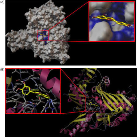
Three-dimensional molecular visualization and structural analysis allow researchers to examine intermolecular distances, hydrogen bonds, hydrophobic contacts, and other molecular features.
Combining these observations with experimental data can improve the interpretation of predicted binding mechanisms.
Factors Influencing Protein–Ligand Binding
Protein–ligand interactions are influenced by multiple physicochemical and structural parameters.
pH and Ionization
The ionization state of amino acid residues and ligand functional groups can change with pH, affecting electrostatic interactions and molecular recognition.
Temperature
Temperature can influence molecular motion, protein flexibility, and the thermodynamic characteristics of binding.
Ionic Strength
Salt concentration affects electrostatic interactions and can influence molecular association and dissociation.
Protein Conformation
Structural changes may modify binding-site accessibility and the spatial arrangement of interacting residues.
Ligand Flexibility
The number of rotatable bonds and conformational freedom of a ligand can influence its ability to adopt a favorable binding geometry.
Solvent Effects
Water molecules and solvent organization can contribute to binding-site interactions and influence the energetic balance of molecular association.
Protein–Ligand Interaction Analysis in Biotechnology
Beyond pharmaceutical research, protein–ligand interactions are relevant to several areas of biotechnology.
In enzyme biotechnology, understanding ligand binding can help investigate substrate recognition and catalytic behavior.
In protein engineering, structural interaction analysis can provide insights into the effects of amino acid substitutions on molecular recognition.
In biochemical research, binding studies can help characterize the interactions between proteins and metabolites, cofactors, or regulatory molecules.
Additional applications include:
Enzyme characterization.
Protein purification research.
Biosensor development.
Biochemical pathway investigation.
Protein engineering.
Molecular diagnostics research.
Biomolecular interaction studies.
These applications demonstrate the importance of molecular recognition in the investigation and development of biological technologies.

Challenges in Protein–Ligand Interaction Studies
Despite significant advances in experimental and computational techniques, investigating protein–ligand interactions remains challenging.
Several factors can complicate the interpretation of molecular binding data.
Protein Flexibility
Proteins are dynamic structures that can undergo conformational changes during ligand recognition.
Static structural models may not fully represent the range of conformations accessible to a protein.
Binding-Site Solvation
Water molecules can participate directly or indirectly in ligand recognition.
Understanding the role of structured water molecules remains an important aspect of molecular interaction analysis.
Computational Prediction Limitations
Docking scores and predicted binding energies are approximations and do not necessarily reproduce experimental binding affinities.
Experimental validation remains important when evaluating computational predictions.
Experimental Conditions
Differences in buffer composition, temperature, pH, protein concentration, and sample preparation can influence measured binding properties.
Careful control of experimental conditions is essential for meaningful comparisons.
Advancing the Understanding of Molecular Interactions
Protein–ligand interactions represent a central subject in molecular and structural biology. Their investigation connects protein architecture, chemical properties, molecular recognition, and biological function.
Through experimental characterization, computational modeling, and structural analysis, researchers can develop a more detailed understanding of the mechanisms governing molecular association.
Continued advances in analytical technologies and computational research will further contribute to the investigation of complex biomolecular systems and the development of innovative approaches in biotechnology.